
Key Takeaways
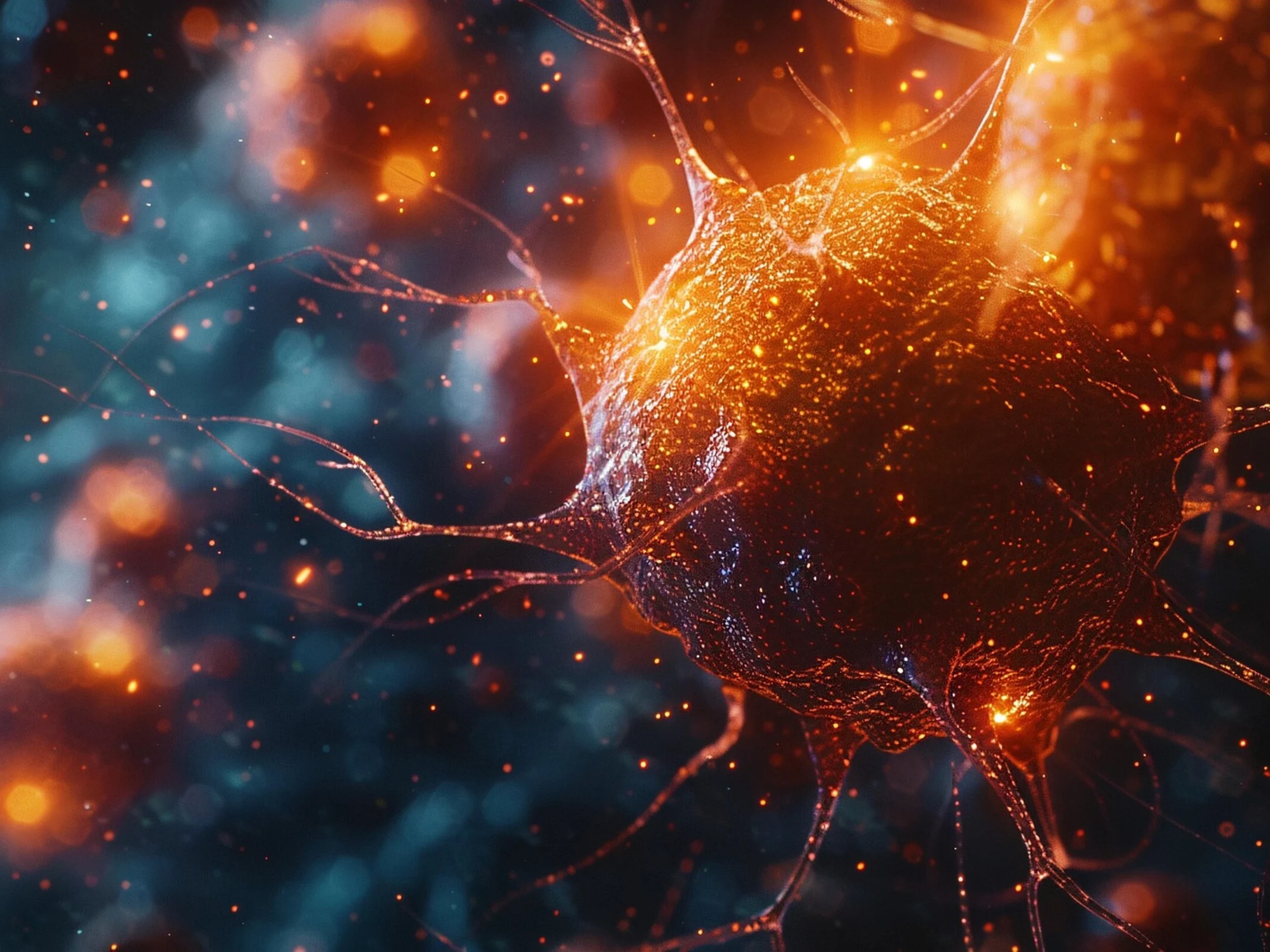
1. The cytoskeleton, made of actin filaments, gives cells their shape and stability, with a newly discovered mechanism for filament breakdown.
2. Researchers identified a collaboration between three proteins—coronin, cofilin, and AIP1—acting together in a “molecular dance” to facilitate actin filament disassembly.
3. Coronin modifies actin filaments, cofilin weakens them, and AIP1 pulls them apart, revealing a new understanding of filament breakdown dynamics.
4. The study challenges previous beliefs by showing that AIP1, not cofilin, plays the key role in severing filaments.
5. Understanding these mechanisms can lead to new therapeutic targets for diseases like cancer, potentially hindering tumor cell movement and metastasis.
Inside each cell, there’s a delicate framework of protein threads called the cytoskeleton, which gives the cell its shape and stability. Actin filaments are essential components of this framework. These small protein strands are constantly being built up and broken down to allow movement. However, the specific mechanism behind how they break down has been a mystery until now.
Discoveries by Researchers
A team from the Max Planck Institute, led by structural biologist Stefan Raunser, has identified that three proteins – coronin, cofilin, and AIP1 – collaborate seamlessly. The researchers liken this collaboration to a “molecular dance,” where each protein has its unique function. Their research was published in Cell in October 2025.
The Role of Each Protein
Initially, coronin attaches to the filament and slightly modifies its structure, facilitating chemical changes, particularly the removal of phosphate groups. This process essentially “matures” the filament, setting it up for what comes next. After that, cofilin takes charge, pushing coronin away and causing further weakening of the filament’s structure. Lastly, AIP1 acts like little pincers, pulling the fragile filament apart and stopping it from being reformed.
To investigate this process, the researchers employed advanced cryo-electron microscopy. This method involves quickly freezing the proteins and using electron beams to create highly detailed 3D images. In total, the team gathered over a million images and pieced together 16 snapshots that illustrate the entire sequence of actions.
Implications of the Findings
The result is a new, detailed model of filament breakdown that challenges previous beliefs. For a long time, cofilin was thought to be the primary protein responsible for severing the filaments. However, it turns out that AIP1 actually holds this key role. The study sheds light on the essential mechanisms that enable cell movement.
These discoveries hold significance beyond basic research. Cell movement is also crucial in various diseases, including cancer and immune responses. In particular, during metastasis – the process where cancer cells spread throughout the body – tumor cells utilize similar mechanisms as healthy cells do when healing wounds.
Now that scientists have a clearer understanding of how proteins like AIP1, cofilin, and coronin regulate cell movement, new targets may arise for therapies. In the long run, this knowledge could lead to treatments that slow down or even stop the spread of cancer cells by disrupting their ability to move.
Leave a Reply